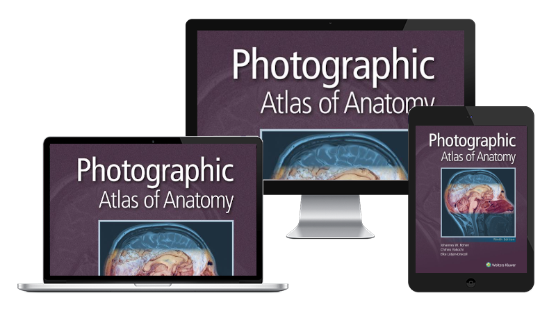
About this edition
Featuring outstanding full-color photographs of actual cadaver dissections with accompanying schematic drawings and diagnostic images, the acclaimed Photographic Atlas of Anatomy, 9th Edition, depicts anatomic structures with unparalleled realism and clarity to help students develop a mastery of human anatomy and excel from the dissection lab to the operating room.
Key features:
- Updated chapters organized by region guide students through the order of a typical dissection.
- A new appendix with learning resources reinforces understanding of the vascular, lymphatic, muscular and nervous systems.
- More than 1,200 full-color dissection photos, medical imaging, and clinical illustrations—all new or updated—depict key anatomical distinctions and functional connections as seen in the dissection lab.
- Functional connections between single organs, the surrounding tissue, and organ systems are clarified to help students prepare for the dissection lab and practical exams.
- Dissections illustrate the regional anatomy in layers “from the outside in.”
- Image bank available for licensing to help support your curriculum.

